Whiplash – Why Does it Happen?

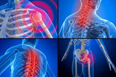
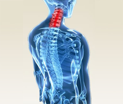
Whiplash injuries are most commonly associated with motor vehicle collisions (MVC), although they can happen from anything that results in a sudden movement of the head—from slip and fall injuries, carnival rides, sports-related injuries, and more. When associated with MVCs, the terms “acceleration/deceleration injury” or “whiplash associated disorders (WAD)” are often applied, depending on the direction of the collision. When the striking vehicle rear-ends the target vehicle, the term “acceleration/deceleration injury” is used. WAD encompasses all scenarios and also includes the type and extent of injury. The degree of injury has been broken down into four main categories with the least amount of injury = WAD I, and the worst soft tissue injury category as WAD III. Fractures are covered separately in the WAD IV category. It has been found that the more severe the soft tissue injury (WAD III > WAD II > WAD I), the worse the prognosis, or the greater the likelihood of long-term injury-related residual problems.

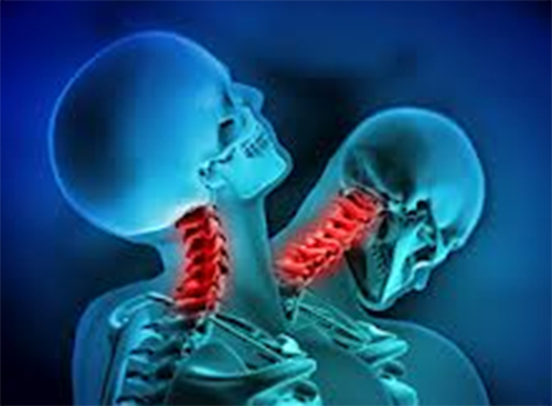
We are often asked why the neck is so vulnerable to injury in a MVC. The simple answer is the head, which weighs about 12-15 pounds (~5-7 kg), is supported by the neck and not all necks have the same length, strength, and mass. This is the reason women (especially those with longer, thin necks) are most vulnerable to the forces that occur in a WAD injury. Another reason whiplash injury can occur is the relatively “slow” speed at which we can voluntarily contract our muscles (>600 msec.) vs. relatively fast speed at which a typical rear-end collision takes to move the head on the neck during whiplash (~300 msec.)! Though the whiplash time duration will vary somewhat, depending on the speed of the collision, angle of the seat back, the distance between the head and the headrest, the “springiness” of the seat back, the weight of the two vehicles, the slipperiness of the road, if the brakes are locked, (…AND MORE!), here’s a typical breakdown of what takes place in a rear-end collision (within a 300 millisecond “typical” time frame):

The degree of injury is affected by all the items previously listed above and more. For example, if the headrest is more than two inches (~5 cm) away from the back of the head, and/or if “ramping” occurs and the head “misses” the headrest, hyper-extension can result and the soft tissues in the front of the neck can become over-stretched and/or the back of the neck can become over-compressed. Or if the rebound phase into flexion exceeds the tissue capacities, the back part of the neck can become over-stretched and the front part over-compressed.

We realize you have a choice in whom you consider for your health care provision and we sincerely appreciate your trust in choosing our service for those needs.  If you, a friend, or family member requires care for Whiplash, we would be honored to render our services.

The Mysteries of Low Back Pain!

Do you realize how complicated the low back region is when it comes to investigating the cause of low back pain (LBP)? There can be findings on an x-ray, MRI, or CT scan such as degenerative disk disease, arthritis, even bulging and/or herniated disks that have NOTHING to do with why the back hurts. Similarly, there are often other abnormal findings present in many of us who have NO low back pain whatsoever! Because of this seemingly paradoxical situation, we as clinicians must be careful not to over-diagnose based on the presence of these “abnormal findings” AND on the same hand, be careful not to under-diagnose them as well.

Looking further into this interesting paradox, one study reported findings that support this point. Investigators examined 67 asymptomatic individuals who hadNO prior history of low back pain and evaluated them using magnetic resonant imaging (MRI). They found 21 of the 67 (31%) had an identifiable disk and/or spinal canal abnormality (which is where the spinal cord and nerves run). Seven years later, this same group of non-suffering individuals were once again contacted to see if they had developed any back problems within that time frame. The goal of the study was to determine if one could “predict” who might develop low back pain based on certain abnormal imaging findings in non-suffering subjects. A questionnaire was sent to each of these individuals, of which 50 completed and returned the questionnaire. A repeat MRI scan was performed on 31 of these subjects, and two neurologists and one orthopedic spine surgeon interpreted the MRI studies using a blinded approach (without having knowledge about the subject’s symptoms or lack thereof). Each level was assessed for abnormalities including disk bulging/herniation and degeneration. Those who had initial abnormal findings were defined as “progressed” (worsened) if an increased severity of the original finding was evident or if additional or new spinal levels had become involved over the seven-year time span.

Of the 50 who returned the questionnaire, 29 (58%) had NO low back pain, while 21 had developed LBP. In the original group that had the MRI repeated seven years later, new MRI findings included the following: twelve remained “normal,” five had herniated disks, three had developed spinal stenosis, and one had “moderate” disk degeneration. Regarding radiating leg pain, four of the eight had abnormal findings originally, two of the eight had spinal stenosis, one had a disk protrusion, and one an “extruded” (“ruptured”) disk. In general, repeat MRI scans revealed a greater frequency of disk herniation, bulging, degeneration, and spinal stenosis compared to the original scans. Those with the longest duration of LBP did NOT have the greatest degree of abnormalities on the original scans. They concluded that the original MRI findings were NOT PREDICTIVE of future development of LBP.

They summarized, “…clinical correlation is essential to determine the importance of abnormalities on MR images.” These findings correlate well with other studies, such as 50% or more of all asymptomatic people HAVE bulging disks and approximately 30% of us have herniated disks – WITHOUT PAIN. To be of diagnostic (clinical) value, the person MUST have signs and symptoms that agree with the imaging test, which is used to CONFIRM the diagnosis. Bottom line, If you have LBP, come see us, as we will evaluate and treat YOU, NOT your x-rays (or MRI) findings!

We realize you have a choice in whom you consider for your health care provision and we sincerely appreciate your trust in choosing our service for those needs.  If you, a friend, or family member requires care for back pain, we would be honored to render our services.

Posture and Headaches

Posture and Headaches (HA) play a significant role in a person’s quality of life and are one of the most common complaints that chiropractors see. This comes as no surprise, as one survey reported 16.6% of adults (18 years and older) suffered from migraines or other severe headaches during the last three months of 2011. Another study reported that head pain was the fifth LEADING CAUSE of emergency department (ED) visits in the United States and accounted for 1.2% of all outpatient visits. These statistics are even worse for females (18-44 years old), where the three month occurrence rate was 26.1% and the third leading cause for ED visits! Because of the significant potential side effects of medications, many headache sufferers turn to non-medication treatment approaches, of which chiropractic is one of the most commonly utilized forms of “complementary and alternative approaches” in the management of tension-type headaches. So, why are headaches so common? Let’s talk about posture!

Posture plays a KEY ROLE in the onset and persistence of cervicogenic headaches. If there is such a thing as “perfect posture,” it might “look” something like this: viewing a person from the front (starting at the feet), the feet would flair slightly outwards symmetrically, the medial longitudinal (inside) arch of the feet would allow enough space for an index finger to creep under to the first joint (and NOT flat like so many), the ankles would line up with the shin bones (and NOT roll inwards), the knees would slightly “knock” inwards and hips would line up squarely with the pelvis. The shoulders would be level, the arms would hang freely and not be pronated (rolled) inwards, and head would be level (not tilted). From the side view, the knees would not be hyperextended nor flexed, the shoulders would not be forward (protracted) and MOST IMPORTANT (at least for headaches), the head would NOT be forward and be able to have a perpendicular line drawn from the floor through the shoulder, as this line should pass through the outer opening of the ear. As the head “translates” or shifts forwards, for every inch of “anterior head translation” (AHT), it essentially gains 10 pounds in weight, which the upper back and neck muscles have to counter balance!

A leading University of California medical author, Dr. Rene Calliet, MD, wrote that this altered posture can add up to 30 pounds of abnormal weight to the neck and can “…pull the entire spine out of alignment.” It can also reduce the lung’s vital capacity by 30%, which can contribute to all sorts of breathing-impaired health problems! Think of carrying a 30-pound watermelon around your neck all day – the muscle pain from fatigue would be tremendous! If this is left uncorrected, chronic neck pain and headaches from pinching off the top three nerves in the neck is likely. The combination of AHT and shoulder protraction may also lead to the development of an upper thoracic “hump” and potentially into a “Dowager Hump” if the Midback vertebrae become compressed (wedged). An increased rate of mortality of 1.44 is reportedly associated with this faulty posture!

Between chiropractic adjustments, posture retraining exercises, other postural corrective care, and strength exercise training, we WILL help you correct your faulty posture so that neck pain and headaches STOP and don’t progress into a chronic, permanent condition.

We realize you have a choice in whom you consider for your health care provision and we sincerely appreciate your trust in choosing our service for those needs.  If you, a friend, or family member requires care for headaches, we would be honored to render our services.

Great Imposters of Carpal Tunnel Syndrome

Carpal Tunnel Syndrome (CTS) is caused by compression and subsequent irritation of the median nerve as it travels through the carpal tunnel and into the hand where it innervates the palm side of the second to fourth digits. As stated last month, the median nerve is sometimes referred to as, “…the eye of the hand” since we rely so heavily on activities of daily living (ADLs) that require its health and function. Some of these ADLs include buttoning a shirt, picking up small objects, tying a shoe or neck tie, writing, holding a book or coffee cup, gripping items such as a phone or steering wheel, opening jars, household chores, and carrying objects, especially with the finger tips.

When patients present with CTS signs and symptoms, one would think that the examination and treatment would be fairly straightforward and “routine.” The problem is, no two cases of CTS are identical because of all the possible mitigating factors, or the presence of OTHER issues that may be contributing or may be the REAL cause for CTS in that particular person. This may explain the reason surgical release of the transverse carpal ligament doesn’t always work!

The “Great Imposters” of CTS include both physical and chemical factors. Physical factors include (but are not limited to): 1) Cervical nerve root compression: Since the median nerve originates from the C6-T1 (and a little from C5) nerve roots exiting the spine, it only makes sense that a pinched nerve in the neck can mimic a pinched nerve at the wrist. The difference here is “usually” that the whole arm is involved, which is less likely in CTS only. Moving down from the neck, the next most common location for a mechanical pinch is at the 2) Thoracic outlet: Here, the nerve roots coming from C5 to T2, like merging lanes of a highway, come together to make the three main nerves that enter the arm and along with the blood vessels, this “neurovascular bundle” leaves the upper chest region and travels through the thoracic outlet to enter into the arm. The thoracic outlet can become narrowed if there is an extra rib, a shift in the collar bone or shoulder blade, from muscles that are too tight (especially the anterior scalene and/or pectoralis minor), or from anything that occupies space within the thoracic outlet. 3) Struther’s ligament: In a few of us (only about 2%), there is a ligament just above the elbow that can entrap the median (as well as the ulnar) nerve, creating a pinch and subsequent numbness below that point, mimicking CTS. 4) Pronator tunnel: The median nerve is more commonly entrapped by the pronator teres muscle just below the elbow, and treating this location can be highly rewarding when managing stubborn CTS cases. Less common is entrapment in the mid-forearm, though it’s possible by either the interosseous membrane that connects the ulna and radius or from fracture of the ulna and/or radius. The most distal point of median nerve compression is at the carpal tunnel. Entrapments can be singular or multiple and when more than one “tunnel” compresses the median nerve, the term double or multiple crush is utilized. Management MUST address ALL points of compression to obtain long-term satisfying results. Other “imposters” of CTS include a host of conditions including (but not limited to) thyroid disease, diabetes, arthritis, pregnancy, birth control pill use, obesity, and MANY others! Chiropractic makes the most sense when it comes to managing CTS from mechanical causes. If response is slow or not satisfying, we will order tests and/or consults to get to the bottom of what “imposters” may be contributing to your CTS symptoms!

We realize you have a choice in whom you consider for your health care provision and we sincerely appreciate your trust in choosing our service for those needs.  If you, a friend, or family member requires care for Carpal Tunnel Syndrome, we would be honored to render our services.

Fibromyalgia: Do I or Don’t I Have It?

Fibromyalgia (FM) is one of the most common types of chronic pain disorders with an estimated five million sufferers in the United States alone. A “hallmark” of FM is the difficulty its sufferers have in describing their symptoms. When asked, “…what type of pain do you feel?,” the response is often delivered with uncertainty such as, “…it’s kind of achy but sometimes gripping…it makes me stop what I’m doing sometimes for only a second or two, but othertimes, I have to sit or lay down until it passes.” It’s sometimes referred to as “deep inside” or radiating, shooting, tender, pins and needles, and locating the pain is another big challenge. It’s often a “generalized” deep ache that includes multiple body areas, sometimes all at once. At other times, it’s spotty and moves around. It’s typically NOT restricted to one side of the body but rather on both sides. It is these inconsistencies that makes diagnosing FM so challenging, sometimes to the point where it can literally takeYEARS before a patient is diagnosed. One study reported that of the 92% FM sufferers who had discussed their complaints with a primary care doctor, only 24% lead to the diagnosis of FM! It is often asked what makes FM so difficult to diagnose and the answer is simply, “…we can’t see it,” and, there are no definitive diagnostics like a blood test, an x-ray, or even more sophisticated tests that can be relied upon to easily make the diagnosis. Moreover, many FM sufferers have other conditions that overshadow FM signs and symptoms that often become the focus of her (or his) doctor.

Back in the early 1990s, the American College of Rheumatology reported “a system” for diagnosing FM. This consisted of a physical examination approach where a certain amount of pressure applied to at least 11 of 18 “tender points” had to be present. This was initially received with enthusiasm, as previously FM was a diagnosis made almost entirely on “gut instinct.” However, it soon became apparent that it was not so easy to interpret the patient’s response when these tender points were tested. Today, for a diagnosis to be made, there are three specific findings that are considered: 1) Wide spread muscle pain (in all four quadrants); 2) Pain that has been present for at least three months; and, 3) at least 11 of the 18 tender points are found – LESS emphasis is placed on the latter. The Fibromyalgia Pain Assessment Tool is a questionnaire filled out by the patient that can also help lead to the diagnosis of FM. Assessing the FM patient for other complaints or conditions commonly associated with FM include the following (% prevalence is reported by fibrocenter.com): 1) Irritable bowel syndrome (32-80%); 2) Temporomandibular disorder (TMD) (75%); 3) Chronic fatigue syndrome – sometimes to the point where bed rest is mandatory (21-80%); 4) Tension or migraine headaches (10-80%); 5) Multiple chemical toxicities; (35-55%); 6) Interstitial cystitis (21%) which includes  eight months of bladder pain, urinary urgency, and frequency (more eight times a day and two times a night); 7) Restless leg syndrome (32%); and 8) Numbness, especially the hands and/or feet (44%). Other common complaints include sleep interference, which prevents deep sleep to be reached, depression or anxiety, concentration and/or memory problems, and more!

As chiropractors, we are trained to assess the FM patient, establish the diagnosis, and offer management strategies such as spinal manipulation, massage, exercise training, nutritional counciling, modalities, and more, which can significantly improve the quality of life of the FM patient. To achieve the best outcome, you may require the services of other types of healthcare providers, as the importance of co-management cannot be overemphasized!

If you, a friend or family member requires care for Fibromyalgia, we sincerely appreciate the trust and confidence shown by choosing our services!

Whiplash Self-Care: Part 2

Last month, we started the discussion of whiplash self-care options in the management of whiplash or CAD (cervical acceleration-deceleration) or WAD (whiplash associated disorders). In this series, we are describing various treatment methods that you can be taught to help facilitate in the management process during the four stages of healing (acute, subacute – discussed last month; remodeling and chronic – addressed this month).

Like in the acute and subacute stages, many of the same self-care techniques can be applied here as well. You will NEVER “hurt” yourself with ice or ice/heat combinations (done properly), so they can be continued indefinitely. Many patients find this helpful. Using the analogy of a cut on the skin, in the acute stage, the cut is fresh and new. It is quite pain sensitive and unstable and it will continue to bleed if you don’t take it easy. After 72 hours (entering the subacute stage), the wound has an immature scab on it and it can still easily be re-injured, and if this occurs, especially by NOT self-managing properly, the recovery time can be significantly prolonged. So, “DON’T PICK AT YOUR CUT!!!” As we enter the later subacute phase (fourteenth week), the wound’s scab is quite mature, and self-care can be appropriately more aggressive. Think strengthening and activity restoration!

Stage 3 – REMODELING phase (14 weeks to 12 months or more): In this stage, we are now three months to a year out from the injury date and hence, we SHOULD now be more “aggressive” with care. During the late acute and subacute stages, you would have been performing exercises focused on movement restoration (range of motion / ROM exercises with LIGHT resistance) in addition to self-applied myofascial release techniques using foam rolls, tennis balls, TheraCane, and/or the Intracell (and possibly others). It is NECESSARY to continue the use of these methods, as they help reduce the chances for any scar tissue to become permanent. In this stage, we will guide you into more advanced exercises that include aerobics (walking, walk/run combinations, etc.) as studies show that whole body aerobic exercise helps MANY specific area injuries, including WAD/CAD injuries. Stretching short/tight muscles, working on balance-challenging exercises (rocker or wobble boards, balance beams, gym balls, eyes closed specific action movements) are VERY IMPORTANT, as they retrain your neuromotor system and reintegrate neural pathways that have been disrupted by the injured tissues and retrain faulty movement patterns you’ve developed from compensating due to pain. Strengthening exercises will include the core since the head sits on the neck, the neck on the trunk, the trunk on the legs, and ALL of this sits on the feet (so we’ll even consider stabilizing the sub-talar joint at the ankle and if pronation is excessive, foot orthotics can help whiplash patients)!

Stage 4: CHRONIC (Permanent): ALL OF THE ABOVE can be employed after the one to two year point to “maintain” your best level of function. If you still have pain, try to “ignore it” and KEEP MOVING, stay active, stay engaged in work, family activities, and DON’T let the condition “win.” AVOID CHRONIC DISABILITY by staying active and fit!

We realize you have a choice in whom you consider for your health care provision and we sincerely appreciate your trust in choosing our service for those needs.  If you, a friend, or family member requires care for Whiplash, we would be honored to render our services.

The “Aging” Lower Back – Part 2

back-painLast month, we started a series on low back pain (LBP) in the geriatric population, and we discussed osteoarthritis (OA) and degenerative disk disease (DDD). As reported last month, this group of conditions often co-exist in this population, so we will continue this discussion this month…

A unique condition associated with OA and DDD is called “spinal stenosis” (SS).  Stenosis means “narrowing,” and it applies to two locations in the spine: 1) The holes through which the nerves in our neck and back exit out of the sides of the spine (called “intervertebral foramen” or, IVF); and, 2) The “spinal canal” through which the spinal cord travels. When narrowing occurs on the sides of the spine where the nerves exit, it’s called, “lateral spinal stenosis.” When the spinal canal narrows, it’s called “central spinal stenosis.” Our spinal cord starts up in the neck as an extension off the brain stem and usually ends at the junction between the middle and lower back (around T12/L1) with the “cauda equina” (which literally means, “horses tail”) and extends downward. The cauda equina is made up of many nerves that travel down and exit out the sides of the lumbar spine (through the IVFs) and sacrum (tail bone) and transfer information (motor and sensory) to and from our legs and brain. When the size of the canal through which these nerves travel close down or narrow enough, sufferers will initially start feeling vague symptoms of leg heaviness or fatigue after walking for 30 or more minutes. As years pass and the IVFs or central canal become gradually more narrow, it may get to the point where a person can only walk a short distance because their legs, “…just won’t move.” A classic complaint of SS is only being able to walk for four to five minutes prior to needing to sit down for 30 seconds to a few minutes (usually five minutes at the most) after which time the leg complaints resolve and the process repeats itself. When the nerves are compressed in these tight canals and the legs become heavy and hard to move, the term, “neurogenic claudication” is used. Another “classic” finding of SS is that RELIEF occurs when the patient bends forward, such as on a grocery cart or, simply stopping and bending over can be immediately relieving in many cases.


Chiropractic adjustments and other techniques are often very helpful in these cases if it is not too far advanced. The good news is that it usually helps, so prior to considering surgery or injections for this, give chiropractic a try – it’s less invasive and safer. We can always refer you to the next step if the condition becomes too advanced and/or if the results become less satisfying.

Compression fractures are another common cause of back pain in the elderly population. They’re often caused by minor trauma in the presence of poor bone density (osteoporosis) which accounts for about 700,000 of the 1.5 million osteoporotic fractures. Interestingly, many patients do not know what they did to cause these fractures so only 25-30% actually go to doctors and have this positively diagnosed (by x-ray). Treatment varies depending on what the percentage of fracture occurred (a little vs. a lot), and in unstable cases, a procedure called kyphoplasty (where cement is injected into the collapsed vertebral body) may be appropriate. As chiropractors, we can help this population by offering nutritional counseling to improve bone density and often provide symptomatic relief with adjustments (low force types) and other modalities.

We realize you have a choice in whom you consider for your health care provision and we sincerely appreciate your trust in choosing our service for those needs.  If you, a friend, or family member requires care for back pain, we would be honored to render our services.


Headaches: How Does Chiropractic Help?

headacheHeadaches (HA) can be tremendously disabling, forcing sufferers away from work or play into a dark, quiet room to minimize any noise and light that intensifies the pain. According to the National Headache Foundation, there are over 45 million Americans who suffer from chronic, re-occurring headaches, of which 28 million are of the migraine variety. Also, approximately 20% of children and adolescents deal with headaches that can interfere significantly with their daily routines. There are many different types of headaches and many sub-types within the main categories. Here are a few: Tension HA (also, called cervicogenic HA), migraine, mixed headache syndrome (a mixture of migraine and tension HAs), cluster (less common but the most severe), sinus headaches, acute headaches, hormone headaches, chronic progressive headaches (traction or inflammatory HAs), and MANY more! Just “GOOGLE” “headache classification” for the daunting list! Let’s take a look at how chiropractic manages these headaches!

According to a study completed in 2005, a review of the published literature revealed good evidence that intensity and frequency of HAs are indeed helped by chiropractic intervention. They limited their review to cervicogenic headaches and spinal manipulation and noted the need for larger scale studies. The well-respected Cochrane database reported spinal manipulation (SM) as an effective treatment option with short-term benefits similar to amitriptyline, a commonly prescribed medication for migraine HA patients.

For cervicogenic HA, the combination of neck exercises and SM was found to be effective in both the short- and long-term, and SM was superior to massage or placebo (sham or “fake” manipulation). Regarding the question of treatment frequency of SM plus up to two modalities (heat and soft tissue therapy), a preliminary study found that when comparing patients receiving one, three, or four visits per week for three weeks, those receiving 9-12 treatments during the three weeks had the most benefit. Regarding the questions, “what is affected by SM” and, “why does SM work” for cervicogenic HA patients, a study describes the intimate relationship between the upper cervical nerve roots (C1-3), the trigeminal (cranial nerve V), the spinal accessory (cranial nerve XI), and the vascular system. Inflammation within these structures and their relationship with the trapezius and SCM muscles help us understand the “why” and “how” of SM and referred pain pattern to the face and head in those with cervicogenic HAs. Realizing this is a bit “technical”, feel free to GOOGLE these structures and you’ll appreciate the close proximity they have to each other and how adjustments, or SM, applied to the upper cervical spine can affect this region. It has also been reported that SM and strengthening of the deep neck flexor muscles benefits the cervicogenic HA patient. Many HA sufferers have combinations of symptoms including dizziness, neck pain, concentration “fog”, fatigue, and others, which were found to also respond to SM applied to the upper cervical spine. One study reported a 36% reduction in pain killer medication use in a group of cervicogenic headache patients receiving SM but no reduction in the patient group receiving soft-tissue therapy. The list of research studies goes on and on! So WHAT are you waiting for? TRY CHIROPRACTIC for your headache management!!!

We realize you have a choice in whom you consider for your health care provision and we sincerely appreciate your trust in choosing our service for those needs.  If you, a friend, or family member requires care for headaches, we would be honored to render our services.

Carpal Tunnel Syndrome – What Is It?

carpal2

Carpal Tunnel Syndrome (CTS) basically occurs when pressure is applied to the median nerve as it travels through the wrist on the palm side resulting in numbness, tingling, pain, and later, weakness of the grip and pinch functions. But, the median nerve can be pinched at many other locations as it courses down from the neck to the hand, which is why we examine and treat the CTS patient from the neck down! The median nerve has been described as the “eye of the hand,” as it is one of the three major nerves formed from the brachial plexus—that “highway” of nerves made up of the C5-T2 roots leaving the neck, merging together to eventually form the three main nerves of the arm. Because the median nerve function regulates pinch and grip strength, buttoning a shirt, writing a note, driving a car, and even sleeping are ALL affected by a median nerve pinch. But WHAT is CTS? Let’s take an “inside” look!

We know that fast, repetitive motion-related jobs like meat or fish packing plants, assembly line work, sewing occupations, and the like can cause CTS over time. Look at the palm side of your wrist and wiggle your fingers. Do you see ALL THE MOVEMENT that is occurring just before the wrist in the forearm? That motion is coming from the tendons, which like shoe strings, attach the forearm muscles to the fingers. Notice ALL the movement in your forearm muscles closer to the elbow – that’s a lot of motion! There are nine tendons that are covered by a lubricating sheath that help the fast moving tendons reduce friction, thus decreasing the chances for heat build up, swelling (inflammation), and subsequent pain and loss of function. But, there is a limit or threshold that the tendons and sheaths can withstand before they just can’t keep up. These nine tendons and sheaths are quite tightly packed together as they leave the forearm and enter the carpal tunnel.

The carpal tunnel is made up of eight small wrist bones called the “carpal bones,” and ANYTHING that makes that tunnel more narrow can effectively cause CTS. If we look at what happens INSIDE the tunnel in the CTS patient, the venous blood flow and nerve flow (called “axonal transport”) is blocked when the PRESSURE inside the tunnel occurs. We all know what it feels like when a blood pressure cuff is inflated on our arm – if it’s pumped up too high or left on too long, the arm REALLY HURTS! That’s because the blood can’t get past the inflated cuff and oxygen can’t get to our muscles and tissues past the cuff and IT CAUSES PAIN!

To give you an appreciation of the pressure difference between the normal vs. CTS wrist, normally, the pressure ranges between 2 and 10 mmHg. We pump up a blood pressure cuff to about 150-200 mmHg when we take blood pressure, so this is NOT MUCH! This 2-10 mmHg pressure increases when we change the position of our fingers, wrist and forearm with wrist extension (bending the hand backwards), causing the greatest pressure increase. This is why we fit the CTS patient with a wrist “cock-up” splint to be worn at night since you can’t control your wrist position when you sleep and any bent position increases the pressure and can wake you up due to numbness, tingling, pain prompting you to shake and flick your hands and fingers until they, “…wake up.” When CTS is present, the pressure inside the tunnel goes up exponentially, meaning NOT 2 or 3 times, but 6, 12, 24 times what is normal and even higher! Now, if you add wrist bending (extension > flexion), the pressure REALLY gets high and it doesn’t take long for the nerve pinch and blood loss to wake us up. We’ve previously talked about other conditions that can make developing CTS more common or make it worse like hypothyroid, diabetes, arthritis, kidney disease, and more. AGAIN, this is because an increase is pressure results from these conditions (increased swelling = increased pressure = increased symptoms). As chiropractors, we will guide and manage your care through the healing process of CTS using a conservative, NON-SURGICAL treatment approach – TRY THIS FIRST!

We realize you have a choice in whom you consider for your health care provision and we sincerely appreciate your trust in choosing our service for those needs.  If you, a friend, or family member requires care for Carpal Tunnel Syndrome, we would be honored to render our services.

Fibromyalgia “(More) Facts”

FibromyalgiaFibromyalgia Facts (FM) has been described as being a “myth” as well as “real” (and probably everything in between the two). This is a VERY controversial disorder that some doctors push under the rug by saying, “….there is no such thing,” while others stake their reputation on it. So with this wide variance in attitude and beliefs about FM, what ARE the facts?

Fibromyalgia has been defined as, “…a complex chronic pain disorder that affects an estimated 10 million Americans” (ref: National Fibromyalgia Association). Women are affected the greatest, but it can affect men and children as well. This condition can be subtle, hardly interfering with life and all of its activities to being totally disabling, disallowing participation in work and the most desired aspects of daily living.

DIAGNOSIS: In 1990, the American College of Rheumatology (ACR) introduced the diagnostic criteria for FM. This includes a patients history of “wide spread pain” for at least three months, AND pain in 11 or more of the 18 specifiic tender points using 4 kg of pressure. Due to the significant controversy about the reality of the disease (as stated in the opening paragraph), ONLY a physician knowledgable about FM should make the diagnosis. Along with this diagnostic responsibility, ALL other conditions having similar presenting symptoms as FM, “…must be ruled out” BEFORE making the diagnosis of FM.

SYMPTOMS: Though the hallmark of FM is widespread, generalized pain (in all four body quadrants), a number of other symptoms are common amongst FM sufferers. Some of these include fatigue (moderate to severe), sleep disorders, brain fog, irritable bowel syndrome (IBS), headaches (including migraine), anxiety, depression, and environmental sensitivities. Studies suggest that there is a “neuroendocrine” (nerves and hormones) abnormality that may contribute to the FM symptoms.

CAUSES: Research has found a genetic link, as FM is OFTEN seen in several family members (among siblings and/or mothers and their children). “Secondary fibromyalgia” arises AFTER other health-related issues occur such as physical trauma (like an acute injury or illness), which can act as a “trigger” for initiating FM. Recently, more attention has been directed to the central nervous system as the “underlying mechanism” for developing FM. Here, the threshold or level of a stimulus that triggers a painful response is found to be much lower in FM patients compared to a healthy group of people (this is called “central sensitization”). Thus, a pain response is amplified in the FM patient due to this lowered threshold of pain tolerance.

TREATMENT: As there is NO KNOWN cure for FM, symptomatic support and functional improvement are two important primary goals when treating patients with FM. In the medical world, there are MANY drugs that have been utilized for FM (such as sleep aids, muscle relaxers, anti-inflammatory, analgesics, and anti-depressants / -anxiety meds). ALTERNATIVE therapies include massage therapy, chiropractic, myofascial release, acupuncture, herbal supplements, yoga, and other exercise approaches such as swimming and/or simply walking are popular care options for many FM patients. Increasing rest, pacing daily activities (to avoid “over-use”), stress management (relaxation tapes, exercise, and nutritional support can ALL HELP reduce FM symptoms and improve quality of life!

If you, a friend or family member requires care for Fibromyalgia, we sincerely appreciate the trust and confidence shown by choosing our services!